What Causes Exercise Intolerance When Initial Heart and Lung Tests Are Normal?
Normal resting tests do not measure every response to exertion
Symptom timing can distinguish breathlessness, orthostatic intolerance, and delayed crashes
Bloodwork, medication review, orthostatic testing, or CPET may identify the next clue
Exercise intolerance with normal heart and lung tests can be frustrating. Patients may wonder, “Why am I exhausted after mild activity?” or experience shortness of breath despite normal tests. Weakness, dizziness, palpitations, or cognitive difficulty may also occur even though an electrocardiogram, echocardiogram, chest X-ray, or pulmonary function test has been described as normal.
A normal result is reassuring, but its meaning depends on which test was performed and whether it measured the body at rest or during exertion. Some problems appear only during activity. Other causes—including anemia, thyroid disease, medication effects, autonomic dysfunction, deconditioning, and post-exertional malaise—may not be detected by routine cardiopulmonary testing.
Why Am I Exhausted After Mild Activity?
Exercise intolerance means being unable to sustain physical activity at a level reasonably expected for a person’s previous function. It is more specific than simply disliking exercise or feeling temporarily out of shape.
Patients may describe:
- Breathlessness that seems disproportionate to the activity
- A rapid, pounding, or unusually slow heart rate
- Lightheadedness, blurred vision, or near-fainting
- Heavy, weak, burning, or painful muscles
- An abrupt loss of stamina compared with prior ability
- Brain fog, headache, flu-like symptoms, or exhaustion after activity
- A delayed crash later the same day or one to two days afterward
The timing matters. Symptoms occurring during activity suggest a different group of possibilities from symptoms that appear primarily after standing, after stopping, or 12 to 48 hours later.
What Do Normal Heart and Lung Tests Actually Rule Out?
“My heart and lungs are normal” may refer to very different evaluations. A resting ECG records electrical activity briefly. An echocardiogram evaluates cardiac structure and function, usually at rest. A chest X-ray does not measure exercise physiology, and spirometry does not exclude every pulmonary or vascular disorder.
These results may lower the likelihood of important diseases without excluding every cardiopulmonary explanation. Intermittent arrhythmias may require ambulatory monitoring, and exercise-induced asthma may require exercise or bronchoprovocation testing. Abnormalities involving cardiac filling pressures, pulmonary circulation, ventilation, or heart-rate response may emerge only during exertion.1,2
The clinician should confirm which tests were completed and whether they were appropriate for the patient’s risks and symptom pattern.
What Causes Exercise Intolerance When Initial Tests Are Normal?
Autonomic dysfunction and orthostatic intolerance
The autonomic nervous system regulates heart rate, blood pressure, sweating, and blood-vessel constriction. If these responses are impaired, standing and movement may reduce effective blood return to the heart or produce an excessive heart-rate response. Symptoms may include dizziness, palpitations, weakness, nausea, brain fog, abnormal sweating, and exercise intolerance.
POTS is one autonomic pattern, but it is not the only one. Orthostatic hypotension, delayed blood-pressure recovery, low blood volume, and other forms of autonomic dysfunction may cause exertional symptoms without meeting POTS criteria. Supine and standing heart rate and blood pressure measurements can provide an initial clue; selected patients may require a standing test or tilt-table test.3,4
Anemia and iron deficiency
Hemoglobin carries oxygen from the lungs to working tissues. Anemia may therefore cause exertional breathlessness, tachycardia, weakness, and reduced stamina even when heart and lung testing is normal. A complete blood count is usually the starting point.
Iron deficiency may contribute to fatigue and impaired physical performance before marked anemia develops. Ferritin must be interpreted in context, and confirmed deficiency should prompt consideration of blood loss, diet, pregnancy, or malabsorption rather than indefinite supplementation alone.5,6
Thyroid, metabolic, and nutritional disorders
Hypothyroidism may produce fatigue, weakness, cold sensitivity, and reduced exercise capacity. Hyperthyroidism can cause heat intolerance, palpitations, weakness, and poor exercise tolerance. Diabetes, electrolyte disturbances, kidney or liver disease, vitamin B12 deficiency, and inadequate nutrition may also limit activity.
Testing should be guided by the history and examination. Common initial studies may include a complete blood count, metabolic panel, thyroid-stimulating hormone, ferritin and iron studies, glucose or hemoglobin A1c, and selected nutritional testing. Broad panels without a clinical rationale often produce incidental abnormalities rather than an explanation.
Medication effects and impaired heart-rate response
Beta blockers may limit the rise in heart rate during exercise. Sedating medications can increase fatigue, diuretics may contribute to volume depletion, and statins occasionally cause muscle symptoms. Other drugs may affect alertness, sweating, or blood pressure.
A heart rate that fails to rise appropriately is called chronotropic incompetence. Medication review and exercise testing may help determine whether medication or sinus-node dysfunction is responsible. Prescribed medication should not be stopped abruptly.
Exercise Intolerance or Just Out of Shape?
Illness, bed rest, pain, or prolonged inactivity can reduce blood volume, strength, and aerobic capacity. Deconditioning usually causes proportional breathlessness and muscle fatigue that improve gradually with carefully progressive activity.
Deconditioning is common, but it should not become a default diagnosis simply because initial tests are normal. A sudden or severe change, fainting, chest discomfort, oxygen desaturation, focal weakness, or delayed multi-day worsening deserves further consideration.
Why Do I Crash After Exercise?
Post-exertional malaise (PEM) is not ordinary tiredness after exercise. It is a worsening of symptoms after physical, cognitive, or emotional exertion that previously would have been tolerated. The decline is often delayed by 12 to 48 hours and may last for days or longer. Fatigue may occur with brain fog, sleep disturbance, pain, headache, sore throat, light sensitivity, or flu-like symptoms.7
PEM is associated with ME/CFS and may occur in Long COVID and other post-infectious illnesses. When this pattern is present, repeatedly pushing through symptoms or automatically prescribing a fixed graded-exercise program may provoke further crashes. Activity management or pacing aims to balance activity and rest within the person’s current limits while the underlying problem is evaluated.7
Sleep disorders, pain, and neuromuscular limitations
Sleep disorders, chronic pain, inflammatory disease, arthritis, and fibromyalgia may reduce physical capacity despite normal resting tests. True muscle weakness—such as difficulty rising from a chair or lifting the arms—may require neurologic examination and targeted testing.
Mitochondrial and metabolic muscle diseases are uncommon. They should be considered selectively when exercise intolerance occurs with objective weakness, cramps, dark urine, an informative family history, or abnormal laboratory findings.
Persistent or post-infectious symptoms
Exercise intolerance may begin during an infection, persist while treatment is underway, or continue after the acute illness. Potential contributors include anemia, autonomic dysfunction, deconditioning, organ injury, medication effects, and post-exertional malaise. The symptom alone cannot determine whether an infection remains active, has resolved, or is unrelated to the current limitation.
This distinction is particularly important for patients with a prior diagnosis of Lyme disease. New or worsening limitation should not automatically be attributed to Lyme disease, but persistent infection also cannot be settled solely by a normal cardiac or pulmonary test. The clinical history, previous treatment, objective findings, and alternative explanations must be considered together. For the Lyme-specific discussion, see Exercise Intolerance in Lyme Disease.
How Does the Symptom Pattern Guide the Evaluation?
- Breathlessness, chest discomfort, or wheezing during exertion: reconsider cardiac ischemia, arrhythmia, exercise-induced bronchoconstriction, pulmonary vascular disease, or dysfunctional breathing.
- Dizziness and palpitations while upright: check orthostatic heart rate and blood pressure and consider autonomic dysfunction or volume depletion.
- Heavy or burning muscles during activity: review anemia, iron status, medications, conditioning, circulation, and neuromuscular findings.
- Delayed worsening 12 to 48 hours later: assess for PEM rather than assuming simple deconditioning.
- Failure of the heart rate to rise—or an excessive rise: review medications, rhythm, sinus-node response, autonomic function, and hydration.
- Progressive loss of function: reopen the evaluation rather than relying indefinitely on earlier normal tests.
When Can Cardiopulmonary Exercise Testing Help?
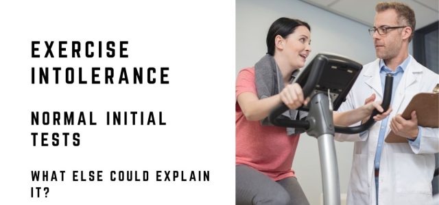
Cardiopulmonary exercise testing (CPET) measures breathing, oxygen use, carbon-dioxide production, heart rate, ECG response, and workload during exercise. It can confirm reduced capacity and help identify whether the dominant limitation appears cardiac, pulmonary, circulatory, muscular, ventilatory, or related to deconditioning. Guidelines consider CPET reasonable for selected patients with unexplained dyspnea.1,2
CPET is not necessary for everyone or definitive in every case. Depending on the symptoms, ambulatory heart monitoring, exercise echocardiography, bronchoprovocation, orthostatic testing, sleep evaluation, or targeted bloodwork may be more appropriate.
A two-day CPET has been used in specialized settings to document impaired recovery in patients with suspected PEM, but deliberately provoking a severe crash carries potential consequences. It should not be treated as routine screening.
When Is Exercise Intolerance an Emergency?
Seek urgent or emergency evaluation when exercise intolerance occurs with:
- Chest pressure, squeezing, or pain
- Fainting during exertion
- Severe or rapidly worsening shortness of breath
- Blue lips, confusion, or low oxygen saturation
- A new sustained rapid or irregular heartbeat
- One-sided weakness, facial droop, or difficulty speaking
- New leg swelling, coughing blood, or sudden pleuritic chest pain
- Dark urine with severe muscle pain or weakness after exertion
Prompt outpatient reassessment is also appropriate when exercise capacity declines substantially, symptoms interfere with ordinary activities, delayed crashes recur, or the condition continues to worsen despite rest and an initial evaluation.
Frequently Asked Questions
What Causes Exercise Intolerance With a Normal Echocardiogram?
Yes. A resting echocardiogram evaluates cardiac structure and function under resting conditions. It may not identify intermittent arrhythmias, an abnormal heart-rate response, exercise-induced filling-pressure changes, or noncardiac causes such as anemia, autonomic dysfunction, medication effects, and post-exertional malaise.
Does normal spirometry rule out a lung-related exercise problem?
No. Normal spirometry is reassuring but does not exclude every respiratory or pulmonary vascular disorder. Exercise-induced bronchoconstriction, impaired gas transfer, dysfunctional breathing, or pulmonary vascular abnormalities may require different testing when the clinical pattern supports it.
How can I tell deconditioning from post-exertional malaise?
Deconditioning usually produces proportional breathlessness and muscle fatigue during activity and improves gradually with carefully progressive conditioning. PEM involves a disproportionate, often delayed worsening of multiple symptoms after exertion and may last for days. The patterns can coexist, so the distinction requires clinical judgment.
Can autonomic dysfunction cause exercise intolerance without POTS?
Yes. POTS is only one form of autonomic dysfunction. Orthostatic hypotension, impaired blood-vessel constriction, abnormal sweating, low circulating volume, and other autonomic abnormalities may limit exercise without meeting POTS heart-rate criteria.
What blood tests may be considered for unexplained exercise intolerance?
Depending on the history and examination, initial testing may include a complete blood count, metabolic panel, thyroid-stimulating hormone, ferritin and iron studies, glucose or hemoglobin A1c, and selected nutritional or inflammatory tests. Testing should be targeted rather than automatically ordering a broad panel.
Clinical Takeaway
Normal initial heart and lung tests are reassuring, but they do not make exercise intolerance imaginary and do not identify every problem that appears during exertion. The most useful next step is to define when symptoms occur, confirm what the initial testing actually assessed, and look selectively at blood counts, iron status, thyroid function, medications, sleep, conditioning, autonomic responses, and post-exertional worsening. CPET or other exercise-based testing may help when symptoms remain unexplained, while urgent warning signs require immediate reassessment.
This article is for educational purposes and does not provide individual medical advice, diagnosis, or treatment. New, severe, or worsening symptoms should be evaluated by a qualified healthcare professional.
Related Articles
- Exercise Intolerance in Lyme Disease
- Autonomic Dysfunction and Lyme Disease
- Lyme Disease and POTS
- What Causes Temperature Intolerance When Thyroid Tests Are Normal?
References
- Guazzi, M., Arena, R., Halle, M., Piepoli, M. F., Myers, J., & Lavie, C. J. (2016). 2016 focused update: Clinical recommendations for cardiopulmonary exercise testing data assessment in specific patient populations. Circulation, 133(24), e694–e711. https://doi.org/10.1161/CIR.0000000000000406
- Heidenreich, P. A., Bozkurt, B., Aguilar, D., et al. (2022). 2022 AHA/ACC/HFSA guideline for the management of heart failure. Circulation, 145(18), e895–e1032. https://doi.org/10.1161/CIR.0000000000001063
- National Institute of Neurological Disorders and Stroke. (2026, March 13). Postural tachycardia syndrome (POTS). https://www.ninds.nih.gov/health-information/disorders/postural-tachycardia-syndrome-pots
- Fu, Q., & Levine, B. D. (2018). Exercise and non-pharmacological treatment of POTS. Autonomic Neuroscience, 215, 20–27. https://doi.org/10.1016/j.autneu.2018.07.001
- Houston, B. L., Hurrie, D., Graham, J., et al. (2018). Efficacy of iron supplementation on fatigue and physical capacity in non-anaemic iron-deficient adults: A systematic review of randomised controlled trials. BMJ Open, 8(4), e019240. https://doi.org/10.1136/bmjopen-2017-019240
- Pasricha, S.-R., Tye-Din, J., Muckenthaler, M. U., & Swinkels, D. W. (2021). Iron deficiency. The Lancet, 397(10270), 233–248. https://doi.org/10.1016/S0140-6736(20)32594-0
- Centers for Disease Control and Prevention. (2024, May 10). Strategies to prevent worsening of symptoms: Treating the most disruptive symptoms first and preventing worsening of symptoms. https://www.cdc.gov/me-cfs/hcp/clinical-care/treating-the-most-disruptive-symptoms-first-and-preventing-worsening-of-symptoms.html
Dr. Daniel Cameron, MD, MPH
Lyme disease clinician with over 30 years of experience and past president of ILADS.
Symptoms • Testing • Coinfections • Recovery • Pediatric • Prevention