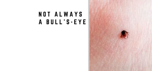
Not Always a Bull’s-Ey
Lyme Science Blog
Sep 11

Lyme Rash Misdiagnosis: Not Always a Bull’s-Eye

3
Visited 5487 Times, 32 Visits today

Lyme Rash Misdiagnosis: Not Always a Bull’s-Eye

No bull’s-eye rash?
A simple red patch instead?
Lyme rashes can be misdiagnosed.

A bull’s-eye rash is often associated with Lyme disease, but the opposite problem is important — Lyme rash misdiagnosis.

Many erythema migrans (EM) rashes do not develop the classic bull’s-eye or ring-within-a-ring appearance. Instead, the rash may be uniformly pink or red, oval, or otherwise different from the familiar textbook image.

A 2022 study by Schotthoefer and colleagues illustrates just how uncommon the classic appearance can be. Two clinicians reviewed photographs from 69 patients with skin lesions suspected to represent early Lyme disease. After review, 35 lesions were classified as erythema migrans. Only 2 of those 35 lesions — 6% — had the classic bull’s-eye or ring-within-a-ring pattern.1

Most of the EM lesions looked quite different. They were commonly uniform, pink, oval, and well-demarcated.1

This matters because patients and clinicians who expect Lyme disease to produce a perfect target-shaped rash may overlook early infection when the skin lesion looks different.

Lyme rashes can also be confused with tick-bite reactions and other skin conditions. In the Schotthoefer study, even a dermatologist and a family physician experienced in recognizing early Lyme disease initially had only moderate agreement when classifying the lesions.1

The belief that Lyme disease always presents with a bull’s-eye is one of the most important Lyme disease misconceptions affecting early recognition.

Clinical Insight: A Lyme disease rash does not have to look like a bull’s-eye. In one study, only 6% of lesions classified as erythema migrans had the classic ring-within-a-ring appearance. Recognizing the broader spectrum of EM may help reduce delayed diagnosis.

How Often Does a Lyme Rash Actually Look Like a Bull’s-Eye?

The classic bull’s-eye is much better known than many of the other appearances of erythema migrans.

In the Schotthoefer study, researchers evaluated 69 people with skin lesions suspected to represent early Lyme disease. Thirty-five lesions were ultimately classified as EM by the two reviewers.1

Among those 35 EM lesions:

  • 6% had the classic bull’s-eye or ring-within-a-ring appearance
  • 51% were uniform in appearance
  • 74% were pink
  • 63% were oval
  • 92% had well-demarcated borders

These findings challenge the idea that central clearing or a target-shaped lesion should be expected before Lyme disease is considered.

Earlier research reached a similar conclusion. Aucott and colleagues showed people photographs of classic and nonclassic Lyme rashes. While 72.7% correctly recognized the classic target lesion, only about 20.5% correctly identified the nonclassic erythema migrans lesions.2

Together, these studies demonstrate an important problem: the Lyme rash people recognize most easily is not necessarily the appearance they will encounter.

What Can an Erythema Migrans Rash Look Like?

Erythema migrans can have a broader range of appearances than the familiar bull’s-eye image suggests.

A Lyme-related rash may appear as:

  1. A uniform pink or red lesion rather than a target-shaped rash
  2. An oval or round expanding lesion without obvious central clearing
  3. Multiple erythema migrans lesions during disseminated infection
  4. A lesion with central darkness or clearing without forming a classic ring-within-a-ring pattern
  5. An atypical lesion that resembles another dermatologic condition

The 2022 study also reinforces why appearance alone may not always settle the diagnosis. Some lesions that were not considered EM were ultimately thought to represent ringworm, allergic contact dermatitis, mosquito or other arthropod bites, hematoma, and other skin conditions.1

Another example is cellulitis. Lyme disease can occasionally produce skin findings that resemble a bacterial skin infection. See Lyme Disease Rash vs Cellulitis: When Lyme Mimics Skin Infection.

Can an Early Lyme Rash Look Like a Tick Bite?

Yes, and this may be one of the most difficult distinctions.

In the Schotthoefer study, the greatest disagreement between the two clinicians involved distinguishing established erythema migrans from possible very early EM and ordinary tick-bite reactions.1

Possible early EM lesions and tick-bite reactions tended to be smaller than established EM lesions. Nearly three-quarters were less than 5 cm. They were also more likely to be round and red and to have a visible central punctum.1

Established EM lesions tended to be larger and had generally been present longer.

Importantly, one lesion initially classified as a tick-bite reaction was associated with subsequent laboratory evidence supporting Lyme disease.1

This illustrates why the evolution of a suspicious skin lesion can matter. A lesion seen shortly after a tick bite may not yet have developed the characteristics that make erythema migrans easier to recognize.

Even Clinicians Can Disagree About Lyme Rashes

One of the most interesting findings from the Schotthoefer study was the degree of disagreement between the two experienced reviewers.

The dermatologist and family physician initially agreed on the classification of only 42 of the 69 lesions, producing moderate inter-reviewer agreement.1

Most of their disagreements involved the boundary between erythema migrans, possible early erythema migrans, and tick-bite reactions.

This does not mean that erythema migrans cannot be recognized clinically. Rather, it demonstrates that very early or atypical lesions may not always fit neatly into a single visual category.

Clinical context—including possible tick exposure, geography, timing, change in the lesion, and accompanying symptoms—can therefore be important when evaluating an unexplained rash.

Can Lyme Tests Be Negative When a Rash Is Present?

Yes. Laboratory testing can be negative during early Lyme disease because the antibody response may not yet be detectable.

The Schotthoefer study illustrates this limitation. Among the 35 patients whose lesions were classified as erythema migrans, only 13 — 37% — had positive laboratory evidence supporting Lyme disease in the study.1

Fourteen of the 35 patients with reviewer-classified EM were negative on all acute serologic tests that were performed.1

This is particularly important because erythema migrans is primarily a clinical diagnosis. A negative early antibody test should therefore be interpreted in the context of the timing of illness and the overall clinical presentation rather than assumed to exclude Lyme disease.

Why Lyme Rash Misdiagnosis Matters

An overlooked erythema migrans rash can represent a missed opportunity to recognize Lyme disease during its early stage.

If a rash is attributed to a spider bite, ringworm, cellulitis, allergic reaction, or another skin condition, Lyme disease may not initially be considered. Patients may subsequently develop additional symptoms as the illness evolves.

These may include fatigue, headache, muscle or joint pain, neurologic symptoms, facial palsy, or joint swelling, depending on the course of the infection.

For a broader discussion, see the Lyme Disease Symptoms Guide.

What Did the Study Not Tell Us?

The Schotthoefer study was small and should not be interpreted as showing that exactly 6% of all Lyme disease rashes are bull’s-eyes.

Only 69 participants were evaluated, and 35 lesions were ultimately classified as erythema migrans. In addition, the participants were almost entirely adults, and all were reported as White and non-Hispanic.1

The findings therefore may not fully represent how erythema migrans appears in children or across different skin tones.

The study nevertheless provides a useful clinical lesson: erythema migrans has a spectrum of appearances, and the classic bull’s-eye represents only one of them.

Clinical Takeaways for Recognizing Lyme Rashes

  1. Do not require a bull’s-eye. Erythema migrans is often uniform rather than target-shaped.
  2. Consider how the lesion changes over time. Very early EM may be difficult to distinguish from a local tick-bite reaction.
  3. Consider the entire clinical picture. Exposure, geography, timing, associated symptoms, and lesion evolution may all contribute to the diagnosis.
  4. Do not use an early negative blood test alone to exclude Lyme disease. Antibody testing can be negative during early infection.

Final Word on Bull’s-Eye Rash Confusion

The assumption that Lyme disease must produce a perfect bull’s-eye can contribute to delayed recognition.

Erythema migrans may instead appear as a uniform pink or red patch, an oval expanding lesion, multiple lesions, or another presentation that does not resemble the familiar target-shaped image.

The 2022 Schotthoefer study makes the point particularly clearly: among 35 lesions classified as erythema migrans, only two had the classic bull’s-eye appearance.1

Recognizing this variability can help patients and clinicians consider Lyme disease even when the rash does not look like the picture commonly associated with the infection.

Frequently Asked Questions

Does a bull’s-eye rash always mean Lyme disease?

No. A target-shaped rash can raise concern for Lyme disease, but other skin conditions can produce ring-shaped lesions. The diagnosis depends on the appearance of the lesion together with the clinical and exposure history.

Can Lyme disease occur without a bull’s-eye rash?

Yes. Erythema migrans frequently does not have the classic bull’s-eye appearance. In one study, only 2 of 35 lesions classified as EM had a classic ring-within-a-ring pattern.

Can a Lyme disease rash be solid red or pink?

Yes. Uniform lesions are common. In the Schotthoefer study, 51% of the lesions classified as EM were uniform and 74% were pink.

Can an early Lyme rash look like a tick-bite reaction?

Yes. Distinguishing very early erythema migrans from a local tick-bite reaction can be difficult. Smaller, round lesions with a central punctum were more common among possible early EM and tick-bite reactions in the Schotthoefer study.

What is a Lyme rash commonly mistaken for?

Erythema migrans may be confused with conditions such as ringworm, cellulitis, allergic or contact dermatitis, arthropod bites, and other skin lesions.

Can Lyme disease testing be negative when erythema migrans is present?

Yes. Antibody testing may be negative early in infection. In the Schotthoefer study, laboratory evidence was found in only 37% of participants whose lesions were classified as erythema migrans.

Should treatment wait for blood tests if erythema migrans is suspected?

Not necessarily. Erythema migrans is primarily a clinical diagnosis, and early antibody testing may be negative. Treatment decisions should consider the clinical presentation, possible exposure, timing, and other relevant findings.

Clinical Takeaway

Lyme rash misdiagnosis can occur because erythema migrans often does not resemble the classic bull’s-eye and because very early lesions can be difficult to distinguish from ordinary tick-bite reactions.

The Schotthoefer study found that only 6% of lesions classified as erythema migrans had a classic ring-within-a-ring appearance. Most were more ordinary-looking pink, uniform, oval lesions.

Lyme disease should remain in the differential diagnosis when a compatible expanding skin lesion develops after possible tick exposure, even when it does not resemble a textbook bull’s-eye.

Related Articles

Atypical Lyme Disease Rash: Cellulitis-Like Lesions and Oral Findings

Lyme Disease Rash vs Cellulitis: When Lyme Mimics Skin Infection

Lyme Disease Symptoms Guide

Why Lyme Disease Gets Missed, Misdiagnosed, or Dismissed

Lyme Disease Misdiagnosis: Why It Happens and What to Know

References

  1. Schotthoefer AM, Green CB, Dempsey G, Horn EJ. The Spectrum of Erythema Migrans in Early Lyme Disease: Can We Improve Its Recognition? Cureus. 2022;14(10):e30673. doi:10.7759/cureus.30673.
  2. Aucott JN, Crowder LA, Yedlin V, Kortte KB. Bull’s-Eye and Nontarget Skin Lesions of Lyme Disease: An Internet Survey of Identification of Erythema Migrans. Dermatol Res Pract. 2012;2012:451727. doi:10.1155/2012/451727.
  3. Dodge T. Lyme Disease: More Than a Bullseye Rash. Adv Emerg Nurs J. 2026;48(1):16-21. doi:10.1097/TME.0000000000000607.

This article is for educational purposes only and is not intended as medical advice. Diagnosis and treatment decisions should be made with a qualified healthcare professional.


Dr. Daniel Cameron, MD, MPH
Lyme disease clinician with over 30 years of experience and past president of ILADS.

SymptomsTestingCoinfectionsRecoveryPediatricPrevention

Related Posts

Leave a Comment

Your email address will not be published. Required fields are marked *