Erythema Migrans Rash: Not Always a Bull’s-Eye
Erythema migrans rash is often not a bull’s-eye
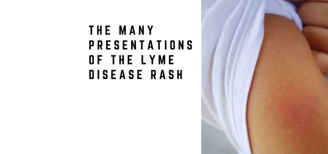
Many Lyme rashes look different than expected
Recognition may improve early diagnosis
In the study “The Spectrum of Erythema Migrans in Early Lyme Disease: Can We Improve Its Recognition?”, investigators examined images of lesions from 69 participants, including 43 men and 26 women, suspected to have early Lyme disease. The majority of participants (83%) presented with a single lesion.¹
The images were retrospectively evaluated by a dermatologist and a family practitioner with expertise in early Lyme disease.
The authors found that 35 lesions (51%) were erythema migrans (EM); 23 lesions (30%) were considered to be possible early EM or tick bite reactions, and 11 (16%) were thought not to be EM, but rather other diagnoses, including ringworm, allergic contact dermatitis, and mosquito bites.
“Only two lesions (6%) were observed with a classic bull’s eye or ring-within-a-ring pattern.”
EM rashes were reported most frequently to appear on the abdomen, thigh, back and hip.
Participants with an EM rash reported the following symptoms: chills, fever, night sweats, headache, fatigue, body aches, nausea and neuralgia.
Because many patients and clinicians still associate Lyme disease with the classic target lesion, recognizing atypical rash patterns is important. Many patients searching for an early Lyme disease rash may not realize how variable these lesions can appear.
What did most erythema migrans rashes look like?
Most EM lesions appeared:
- Uniform (51%)
- Pink (74%)
- With an oval shape (63%)
- Well-defined borders (92%)
Many erythema migrans lesions appear solid and expanding rather than ring-shaped.
What did early EM or tick bite reactions look like?
They “were typically <5 cm in size (74%), red (52%), round lesions (61%), with a punctum present (100%),” according to the authors.
Distinguishing an early EM rash from a tick bite reaction can be difficult. Patients with recent bites may also benefit from reviewing bad signs after a tick bite.
What did lesions that were not EM look like?
Lesions that were not EM rashes appeared pink or red (64%), round (55%), or uniform (45%) lesions, but also had raised (25%) or irregular borders (33%).
These findings show why Lyme rashes are frequently confused with fungal infections, allergic reactions, and other skin conditions. This overlap contributes to Lyme disease rash misdiagnosis.
“EM commonly occurs in forms that are not the classic bull’s eye.”
“Only 14 (20%) participants overall had positive laboratory evidence for LD; these included 13 (37%) of the participants with EM-classified lesions,” the authors wrote.
An expanding rash accompanied by fatigue, headache, fever, or neurologic symptoms may increase suspicion for early Lyme disease.
The authors suggest that “education should deemphasize the bull’s eye form and stress the wide variability in EM instead and the fact that many of them present as a uniform, homogeneous lesion.”
Why recognition matters
This matters because early Lyme disease is often diagnosed clinically, especially when an expanding rash appears after possible exposure. Testing may be negative early, making rash recognition especially important. For broader diagnostic context, see Lyme test accuracy.
Because many patients do not recall a tick bite, rash recognition should be considered alongside exposure risk, symptoms, and timing after outdoor activity. Patients concerned about exposure may also review Lyme disease prevention.
The authors conclude:
- “Patients often present with lesions that may represent the very early stage of EM or tick bite reactions, and most patients will test negative on currently available laboratory tests…”
- “Clinicians may not be aware of all existing variations, such that some LD patients with EM may not be immediately recognized and promptly diagnosed and treated. Hence, further improvements in terms of clinician awareness and recognition of EM are needed.”
Frequently Asked Questions
Does an erythema migrans rash always look like a bull’s-eye?
No. Only 6% of lesions in this study had the classic bull’s-eye appearance.
What is the most common appearance of an erythema migrans rash?
Many lesions appeared uniform, pink, oval-shaped, and well-defined rather than ring-shaped.
How soon does an erythema migrans rash appear?
An erythema migrans rash often appears days to weeks after a tick bite, though timing varies.
Can Lyme disease testing be negative when an EM rash is present?
Yes. Early Lyme disease testing may be negative even when an EM rash is present.
Can an early Lyme rash be mistaken for a tick bite reaction?
Yes. Small lesions and early EM may overlap visually with local bite reactions.
Clinical Takeaway
An erythema migrans rash does not always look like a bull’s-eye.
Many lesions appear uniform, pink, expanding, or oval-shaped rather than ring-shaped.
Recognizing the many presentations of erythema migrans may reduce missed diagnoses and improve early treatment decisions.
Related Articles
What does a Lyme disease rash look like?
Lyme disease rash puzzles doctors, leads to misdiagnoses
Erythema migrans rash doesn’t always have Bull’s-eye appearance
Is it too late to treat a tick bite?
References
- Schotthoefer AM, Green CB, Dempsey G, et al. The Spectrum of Erythema Migrans in Early Lyme Disease: Can We Improve Its Recognition? Cureus. 2022;14(10):e30673. doi:10.7759/cureus.30673.
Dr. Daniel Cameron, MD, MPH
Lyme disease clinician with over 30 years of experience and past president of ILADS.
Symptoms • Testing • Coinfections • Recovery • Pediatric • Prevention


My rash was a huge hour glass shape on my whole torso region. My primary thought it was a fungus and was going to give me a fungicide. I thought better of it and told him it was Lyme related and to test me for Lyme which was confirmed.
I am your doctor was willing to work with you.
Can lyme or any of it’s co-infections cause ones A1c to climb to Diabetic numbers?
There are so many causes for HA1C. I have HA1C rise in some Lyme disease patients who have cut out activity, gained weight, and were not able to follow a diet.