Can Lyme Disease Show Up on an MRI? What Brain Imaging Reveals
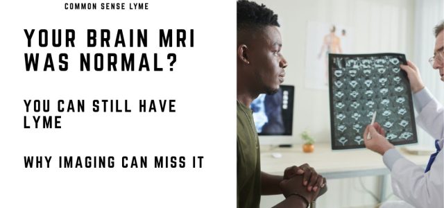
Can Lyme disease show up on an MRI? Sometimes—but often it does not.
Many patients with neurologic Lyme disease have normal brain imaging, even when symptoms such as brain fog, memory problems, or nerve pain are severe.
Quick answer: Brain MRI in Lyme disease may show abnormalities, but findings are often nonspecific. A normal MRI does not rule out neurologic Lyme disease.
For a broader overview, see Lyme disease symptoms guide.
Brain MRI and Neurologic Lyme Disease
Neurologic Lyme disease can affect the brain, spinal cord, and peripheral nerves.
MRI is often used when patients present with:
- Memory problems or cognitive decline
- Facial palsy
- Neuropathy
- Headache or meningitis-like symptoms
In some cases, MRI may show abnormalities that help clinicians evaluate neurologic symptoms.
What MRI May Show in Lyme Disease
Some patients develop white matter changes on MRI.
These may appear as multiple areas of T2 hyperintensity in the brain, particularly in periventricular or subcortical regions.
These findings can resemble other neurologic conditions such as multiple sclerosis.
Important: MRI findings alone cannot confirm Lyme disease.
Functional Imaging: When MRI Is Normal
Structural imaging such as MRI may appear normal, even when symptoms are significant.
Functional imaging, such as FDG-PET scans, has identified patterns of reduced brain activity in some Lyme patients, including:
- Temporal lobe hypometabolism
- Diffuse cortical hypometabolism
These regions are involved in memory and cognition, helping explain symptoms such as brain fog and slowed thinking.
While FDG-PET imaging is not routinely used to diagnose Lyme disease, it may reveal functional brain changes in selected patients with neurologic involvement.
Facial Nerve and Cranial Nerve Findings
MRI may also detect abnormalities in cranial nerves.
In Lyme-related facial palsy, imaging may show nerve enhancement, though this finding is not specific to Lyme disease.
Lyme facial paralysis can resemble Bell’s palsy or other causes of nerve dysfunction.
MRI may also show enhancement of the meninges or spinal nerves in patients with meningoradiculitis.
Radiologic imaging can sometimes help clinicians distinguish between direct neurologic involvement and secondary encephalopathy.
Why a Normal MRI Does Not Rule Out Lyme Disease
This is the most important point.
Many patients with neurologic Lyme disease have completely normal MRI scans.
CT and MRI imaging may fail to detect infection-related changes, particularly early or in cases involving small fiber or functional dysfunction.
This is why diagnosis relies on:
- Symptom patterns
- Exposure history
- Laboratory testing
Learn more about why Lyme tests can be negative.
Frequently Asked Questions
Can Lyme disease show up on MRI?
Sometimes, but findings are often nonspecific. Many patients have normal imaging.
What does Lyme disease look like on MRI?
It may show white matter lesions or nerve enhancement, but these findings are not unique to Lyme disease.
Can you have neurologic Lyme with a normal MRI?
Yes. A normal MRI does not rule out neurologic Lyme disease.
Can Lyme disease brain fog happen with a normal MRI?
Yes. Many patients with brain fog or cognitive symptoms have normal structural imaging despite significant neurologic symptoms.
Clinical Perspective and Takeaway
Normal brain imaging does not rule out neurologic Lyme disease.
Patients with brain fog, neuropathy, cognitive symptoms, or facial palsy may still have neurologic involvement even when MRI findings are normal or nonspecific.
Imaging must be interpreted alongside symptoms, exposure history, and laboratory evaluation.
Related Articles
- Brain Fog in Lyme Disease
- Lyme Disease Facial Palsy
- Neurological Damage and Dysfunction in Early Lyme Disease
- When It Looks Like a Brain Tumor but It Is Lyme Disease
References
- Alves Simão AK, Amaral LLFD, Inada BSY, et al. Neuroimaging of Emergent and Reemergent Infections. Radiographics. 2019;39(6):1649-1671.
- Fallon BA, Nields JA. Lyme disease: a neuropsychiatric illness. Am J Psychiatry. 1994;151(11):1571-1583.
- Newberg A, Hassan A, Alavi A. Cerebral metabolic changes associated with Lyme disease. Nucl Med Commun. 2000;21(8):773-777.
Dr. Daniel Cameron, MD, MPH
Lyme disease clinician with over 30 years of experience and past president of ILADS.
Symptoms • Testing • Coinfections • Recovery • Pediatric • Prevention


Hi Dr Cameron, Was wondering can the findings of Microvascular ischemic and changes of white mater be caused by Lyme? I had a CT scan yesterday no contrast and a spinal tap no results on that yet it was compared to a CT from 6 months ago. I was straight positive on Lyme Elisa and Western blot nov 2021 may have had it sinse 2016 didn’t get right treatment then over the past summer Western blot only lost one of the bottom bands now that band is back been told I have active infection but don’t think 100mg doxy 2x a day and ivermectin is going to be enough trying to deal with Cleveland clinic Dr’s for the sever neurological symptoms is tough as they believe one treatment is enough! I’m currently being treated by ILADS Cnp but I’m not getting better .. Also had Babesia duncani 160 IgG IgeneX and a positive Bartonella from Mdl that said I was positive for 3 different species of Lyme.. Can Lyme cause these findings ? Having horrible neurological issues, nervousness, insomnia, emotional feeling of impending doom and confusion..
Thank you,
Jim
White spots on an MRI are from so many causes. I advise my patients to be reassessed for tick borne illness including Babesia.
Hey Jim,
I was diagnosed with Lyme a few weeks back and am also on a journey to figure all of this stuff out. I read your post and my heart went out to you as the last few years I’ve had the same horrible symptoms. It really is awful to feel so anxious, confused, and dark all the time! Especially when you have no idea why and you think you’re losing your mind (which is how it was for me since is didn’t know about the Lyme yet).
I just wanted to reach out and say you’re not alone on this tough, tough journey. I’m hoping for the best for you. ❤️ ✨
Hi Jim. I am wondering how you are making out. What treatments you’ve received. My husband currently is hospitalized in a BSU with paranoia and hallucinations…which all stemmed from insomnia, and I believe directly from Lyme. Awaiting results. He also has “mild small vessel ischemic areas” on his MRI. Thanks so much for your response.
God morning, When a doctor (Dr. Andrew Serpe, Amityville) finally figured out my problem with a blood test it was discovered that I had Lyme disease. How long I do not know. But my blood work showed 7 bars effected by lyme. I am now on gabapentin 300mg 3 times a day. I really don’t believe in or want to take drugs. But the numbing and pain in my feet is getting worse . If I had to guess I was bite 20-25 years before diagnoses, which was 3 years ago. Is this like diabetes where I will be loosing my toes ?? If you can not help me can you recommend someone ?? I hope you will read this and help me. I am a 70 year old very active women. Thank you for your kindness Jacqueline Smith
Numbness can occur in Lyme and diabetes. I have had patients in my practice with both.
I had a huge tick in ear for 9 months when found doc through it away,I have had 4 MRI done and no one saw it? I’m sooo sick and waiting for results.im positive it’s lymes
The test are not as reliable as I would like. I have had to treat using clinical judgement if the tests are negative.
Hi dr. Cameron. My husband is 43 years old, no history of mental health issues, and over the past week, he has had increasing paranoia, and started hallucinating. MRI revealed “ BRAIN PARENCHYMA: There are scattered foci of signal abnormality in a
bifrontal distribution suggestive of sequelae of prior
headaches/migraines or early chronic small vessel ischemic disease.”. Doctor sent out Lymes panels and not back yet. Do you have any insight on this? Any help is greatly appreciated. I need my husband back. Thanks so much.
Hemp Dr Cameron. Was wondering about the effects of tick borne disease on having flares in the T2 area? Can you explain this maybe? Thanks
Tick-borne illness can involve neurologic and musculoskeletal symptoms, but focal flares need clinical evaluation rather than assumptions.